When you slip a slide under a microscope, a system of glass lenses magnifies the object of your attention — a microbe, for example. But even with the largest zoom on a classic compound optical system, scientists struggle to make sense of finer details, which can be further obscured when tough cell walls make it difficult to inject dyes that help identify structures.
Now, rather than invest in more powerful and more expensive technologies, some scientists are using an alternative technique called expansion microscopy, which inflates the subject using the same moisture-absorbing material found in diapers.
“It’s cheap, it’s easy to learn, and indeed, on a cheap microscope, it gives you better images,” said Omaya Dudin, a cell biologist at the University of Geneva who studies multicellularity.
Expansion microscopy was developed by Ed Boyden at the McGovern Institute for Brain Research at the Massachusetts Institute of Technology in 2015. Boyden and colleagues successfully expanded a biological sample by infusing it with a hydrogel made of sodium acrylate. A key ingredient layered in diapers to keep babies dry, the compound can absorb hundreds of times its weight in water while retaining its overall structure.
In expansion microscopy, specific biomolecules such as proteins are anchored to the gel. As the gel absorbs added water, its weblike matrix swells, and the space between the web’s anchor points dilates. Ideally, the overall structure remains, allowing researchers to visualize extra-tiny anatomy or see inside cells with tough barriers.
Dudin had spent six frustrating years trying to force antibodies through his target cells’ sturdy walls to bind to specific proteins and visualize their internal structures, and he was only able to do so through a complex freeze-and-thaw protocol that destroyed most of the final product. Desperate, he struck up a Covid-era collaboration with the lab next door that was using expansion microscopy.
“That moment was just magical. All the cells were expanded, everything stained, we could see,” Dudin said. “It very rapidly became clear that we should aim for the sky with this one.”
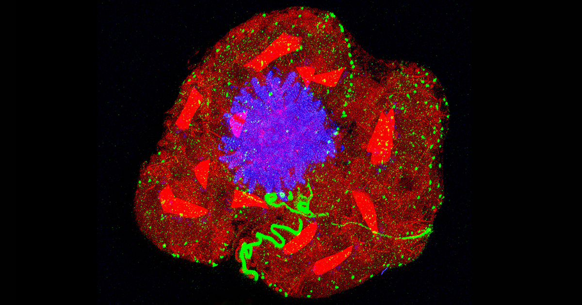
Gautam Dey, a cell biologist at the European Molecular Biology Laboratory in Heidelberg who studies mitosis, found that the method worked just as well in his lab. The samples were clearer, and the dyes and antibodies penetrated cells more effectively, so the two labs struck up a collaboration to visualize species they had never studied before. They are working to chart the landscape of cytoskeletal diversity, visualizing complex cytoskeletal structures that have never been seen in such detail.
Perhaps most importantly, expansion microscopy is possible for any lab with a basic microscope and the hydrogel. “People have talked about democratizing microscopy before. This is it, it’s happening,” Dey said. “I think it’s just a matter of time before any cell biology lab in the world is doing it. A basic fluorescence microscope is never too far away.”